Computed tomography scan (CT)
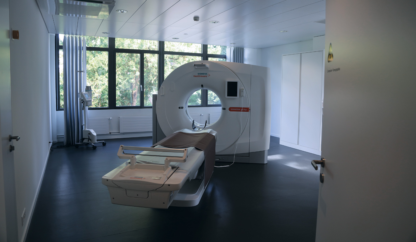
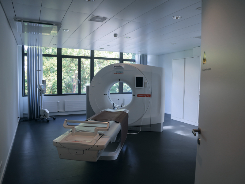
Computed tomography scan (CT) is a fast method that uses X-rays to show the density of the organs under examination. It allows fine separation of both calcified and ossified structures. With the new generation of CT machines (Siemens Somatom go.up), it is also possible to perform dynamic examinations, to visualize vessels by means of CT angiography or to visualize cerebral blood flow by means of perfusion CT. Interventions can also be guided by CT, e.g. in pain therapies.
Examination procedure
Since the CT is a large rotating X-ray tube, a protective jacket may be necessary. During registration and planning, our secretary will clarify everything necessary with you and make the necessary preparations for the examination together with you.
Immediately before the examination, you will receive an information sheet and questionnaire from us, which our radiology specialists (MTRA) will go through with you.
For some examinations, a contrast medium containing iodine is necessary. This allows vessels to be visualized and tissues, such as the vessel wall, to be better differentiated. The contrast medium is usually very well tolerated. If you suffer from allergies or have had previous contrast agent intolerances, please inform us in good time.
CT in brief:
- sometimes a contrast medium is necessary
- as in a normal X-ray examination, X-rays are also used in a CT examination
- you wear special clothing
- we see and instruct you
- you can talk to us at any time
The examination may require you to wear special clothing. After you have changed, you will be prepared for the examination by our MTRA. Our latest generation Siemens CT has a large tube opening and a comfortable couch where the MTRA will position you for the exam. Our MTRA can see you and communicate with you via an intercom system. If you feel uncomfortable, you can use a bell to make yourself known.
For optimal image acquisition, it is important that you do not move throughout the examination. It is best to keep your eyes closed during head exams.
After the examination, your images will be post-processed by MTRA and then analyzed and evaluated by the radiologist. The images are stored in our findings portal for 15 years in accordance with data protection guidelines. You can access them there at any time.